Science really gets me excited. What is most exciting is when something new is discovered. This could be a new treatment, new technology, or revisiting of a previously thought notion or idea.
Eyes are one of the coolest organs in the body. They are windows to the brain and some would say to the soul. In humans the vital sign of the eye is visual acuity—you know, when you have to cover one eye and read the eye chart. 20/20 vision means that you see the same as a normal eye would see at 20 feet. 20/100 vision means that you see what a normal eye would see at 100 feet. As you might imagine it’s much more difficult to test the visual acuity of a dog’s eye—they can’t tell us what they see. Due to this, some scientists believe that the visual acuity of the dog has been underestimated.
We know that dogs see differently than humans do. For many years we have known that dogs can see much better in low light than us; they have rapid vision that allows them to detect rapid changes in the light, and, due to the placement of their eyes in their skulls, they have wider visual fields than we do (Miller & Murphy, 1995).
All of these qualities aid the dog, a predator, in its ability to hunt. However, the acuity at which the dog can focus was thought to be diminished, as the dog is known to lack a fovea (Miller & Murphy, 1995). Fovea centralis (fovea) is a structure in the human eye. The fovea is a depression within the retina that contains a large number of densely packed cones type cells that are responsible for visual acuity (Beltran et al., 2014).

For years scientists have felt that the dog’s visual streak was responsible for their visual acuity. The visual streak is an area in the retina with increased amounts of photosensitive retinal ganglionic cells and cone cells (Miller & Murphy, 1995). However, in 2014 things changed.
In 2014 the canine retina was evaluated with in vivo (in life) and ex vivo (in death) imaging (Beltran et al., 2014). The researchers found an area in the retina very similar to a non-human primate fovea, which they deemed the area centralis. This area was tiny but full of densely packed cone cells (Beltran et al., 2014). This area was not a fovea-like depression but was very similar from a histologic standpoint to what is seen in the center of the human fovea, the foveola (Beltran et al., 2014).
You may be asking why is she so excited about this? This information is incredibly important. This indicates that a dog’s visual acuity is actually better than previously thought. The visual acuity of a dog was thought to be about 20/50 (Miller & Murphy, 1995). Based on the findings in this study, the visual acuity of the dog would be between 20/24 and 20/13 (Beltran et al., 2014). That means that dogs could be able to see at 20 feet what a normal eye would see at 13 feet!
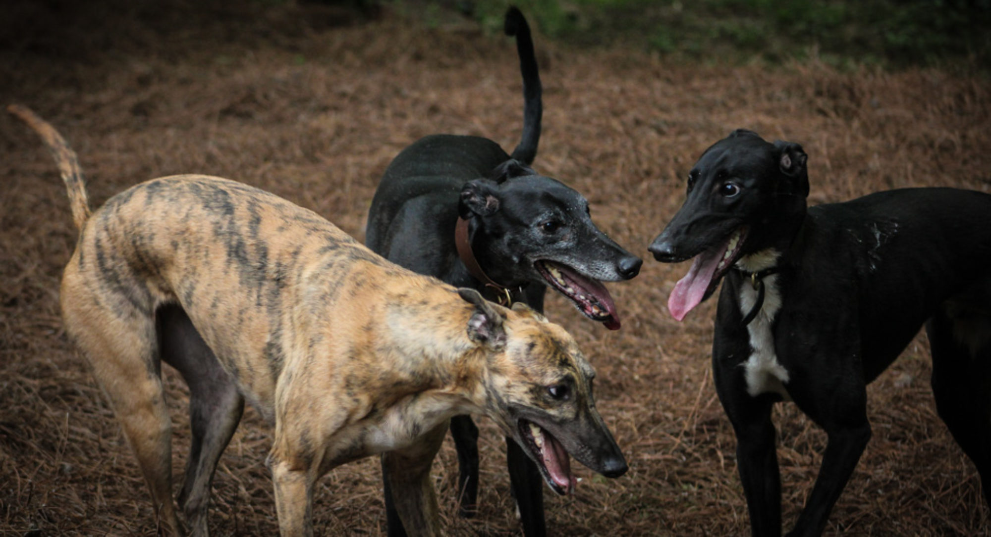
The canine eye is an important structure for multiple reasons. For our Greyhounds the eye is important for racing, lure coursing, coursing, hunting, fetching, running through agility obstacles, and their everyday lives. Just imagine having better than perfect vision and then adding a wider visual field, the ability to detect rapid changes in light, and the ability to see in low light—I would be overwhelmed with that much stimuli going through my brain all the time! Dogs are complex animals and the more we learn about them the more I amazed by all they do!

Beltran, W. A., Cideciyan, A. V., Guziewicz, K. E., Iwabe, S., Swider, M., Scott, E. M., Aguirre, G. D. (2014). Canine retina has a primate fovea-like bouquet of cone photoreceptors which is affected by inherited macular degenerations. PLoS One, 9(3), e90390. doi:10.1371/journal.pone.0090390
Miller, P. E., & Murphy, C. J. (1995). Vision in dogs. J Am Vet Med Assoc, 207(12), 1623-1634.

